For most patients, symptoms and clinical presentation may proceed diagnosis. Patients with GCT of the bone often present with bone pain, swelling, tenderness, range of motion difficulties, and pathological fractures. This presentation is seen in other diseases, such as, aneurysmal bone cyst, telangiectatic osteosarcoma, brown tumor of hyperparathyroidism, and malignant fibrous histiocytoma (1,2). The symptoms and osteoclasts associated with GCT of the bone are common in other diseases, making a differential diagnosis necessary for GCT of the bone patients. A differential diagnosis means that the healthcare provider ruled out other possible diseases which led to the symptoms and pathology.
Therefore, biopsy is often required to exclude other potential diseases prior to diagnosis of GCT of the bone. To learn more about biopsies, go to Biopsy.
However, symptoms can vary widely among patients, especially dependent on grade and location of the GCT of the bone. Patients with Grade III GCT of the bone are more likely to exhibit severe symptoms and pathological fractures, while Grade I GCT of the bone patients may have no symptoms. This may not always be the case. In some cases, Grade I GCT of the bone patients may present with mild symptoms, there are many factors that play a role in symptom onset. The presentation of this disease is hard to predict and cannot be predicted solely off imaging or biological features. Therefore, patients medical history and physical examination are crucial for the right diagnosis.
Imaging modality
The most common imaging modalities used in the diagnosis of GCT of the bone are radiographs. Each imaging technique provides strengths and weaknesses depending on the intent.
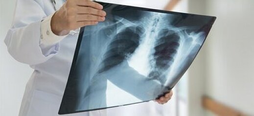
X-RAY
In daily life, the most common radiology technique is radiography. Radiography uses x-rays to view internal structures of the body, mostly skeletal. X-rays use a type of radiation known as electromagnetic waves which will produce an image on metal film. Soft tissue, such as skin and organs, cannot absorb the electromagnetic waves and allows the high energy to pass through them. Once the high energy waves reach bone and other solid structures, the radiation will be absorbed allowing an image of those regions. X-rays are the least expensive of the imaging techniques and generally the quickest. Degeneration, effusion, bone erosion, and the amount of joint space can be identified on x-ray, giving a quick snapshot of what may be going on internally. Due to the location of the tumor occurring in bone, typically x-rays provide a clear indication of the bone health and the degree of bone decay caused by the tumor. This is the most common imaging modality used in GCT of the bone, especially following pathological fracture.
Additionally, it is often necessary to do chest x-rays every 6-12 months to monitor for lung metastasis depending on the patient. Consult with your healthcare provider about monitoring for metastasis.
Computed tomography scan (ct scan)
Computed Tomography, commonly referred to as a CT scan, is an imaging technique that utilizes a combination of x-rays and a computer to create a picture of organs, bone, and other tissues. A series of images are taken using x-rays at different angles around the body, giving 3 dimensional information. This technique allows for more accuracy than a x-ray. This is commonly done prior to surgery to enhance the quality of what is known about the tumor.
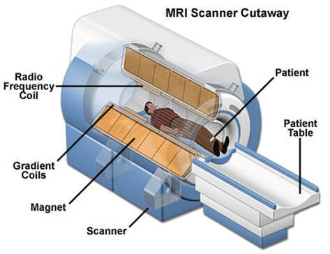
Magnetic resonance imaging (mri)
Magnetic resonance imaging (MRI) allows for soft tissue to be visualized in the most clear and optimal way. A MRI is an imaging technique that utilizes a strong magnetic field and radio waves to generate an image that cannot be seen well on x-rays. MRI does not use radiation, instead, it relies on atoms in your body to align with the moving magnets in the scanner. During a MRI scan, radiofrequencies are pulsed through a patient allowing their atoms to move according to the magnets. This alignment with the magnets leads to an image shown in T1 and T2 intervals. The joint can be easily depicted during an MRI. T2 intervals refer to the appearance of fluid, often in this case, synovial fluid and effusions. GCT patients often have swelling leading to excess T2 intervals present. GCT tumors require a blood supply to receive nutrients and grow. Because of this blood supply to the tumor, they also show up on T1 intervals. This allows more sophisticated information to be gained about the tumor. Often times the MRI is coupled with contrasting dye. The contrasting dye injected, either locally via arthrogram or through an intravenous injection, is a gadolinium-based dye that enhances the quality of the imaging. This allows radiologists to more confidently identify soft tissue tumors. The contrasting dye temporarily enhances the T1 intervals, leading to more accurate diagnostics. Specifically, contrast agent is added to scans to visualize tumors, inflammation, certain organ's blood supply, and blood vessels. Not every MRI will require contrasting dye. Your healthcare provider will determine whether to add contrast or not based on your present condition and your health history.
Often times, GCT of the bone may be clear using radiography alone and may not require an MRI. Consult with your healthcare provider for the proper imaging modality.